

Differentiation of Stem Cells at Future Health
One of the defining characteristics of stem cells is their ability to turn into other cell types in the body, allowing the body to use these cells to repair and regenerate tissues.
This ability is known as plasticity.
The process of stem cells turning into other cell types is called differentiation. It occurs naturally within the body and starts at the very beginning of life – you start as a fertilised egg cell which divides into 2 cells, then 4, then 8, then 16 and so on. After a while, these cells start turning into other cell types and eventually they will go on to form all the cell types that make up the human body (nerve cells, muscle cells, blood cells, skin cells etc).
The Human Tissue Authority require us to perform many experiments to demonstrate that the samples we store at Future Health fulfil all sorts of quality criteria, demonstrating that they are of suitable quality for potential future therapeutic use. This includes demonstrating that the cells we store from cord tissue and dental pulp can be differentiated.
To do this, we take cord tissue and dental pulp stem cells and culture them in the lab with various chemical cocktails that are designed to make them turn into other cell types.
The standard method to demonstrate that cells have retained their plasticity is to turn them into osteocytes (bone-forming cells), chondrocytes (cartilage-forming cells) and adipocytes (fat-forming cells).
This process is known as tri-lineage differentiation. From starting the culture through to imaging the final result takes around 5-6 weeks.
Below are a few pictures (taken down a microscope) from some recent experiments.
To give an idea of scale, the bar in the lower right corner of the pictures is a tenth of a millimetre long.
The first picture shows some undifferentiated cells at the start of the experiment.

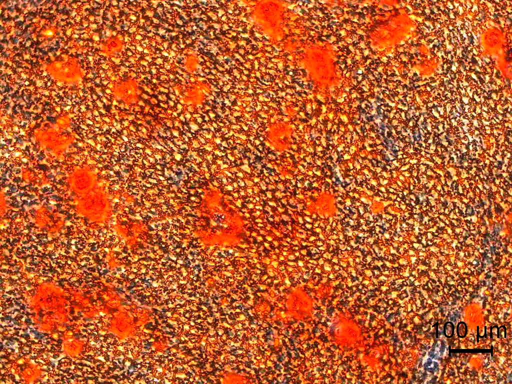
This picture shows dental pulp cells which have been turned into osteocytes (bone-forming cells).
To demonstrate that the differentiation has been successful, the cells are stained with a dye called alizarin red which specifically stains calcium deposits (Calcium, of course, being a major component of bone).
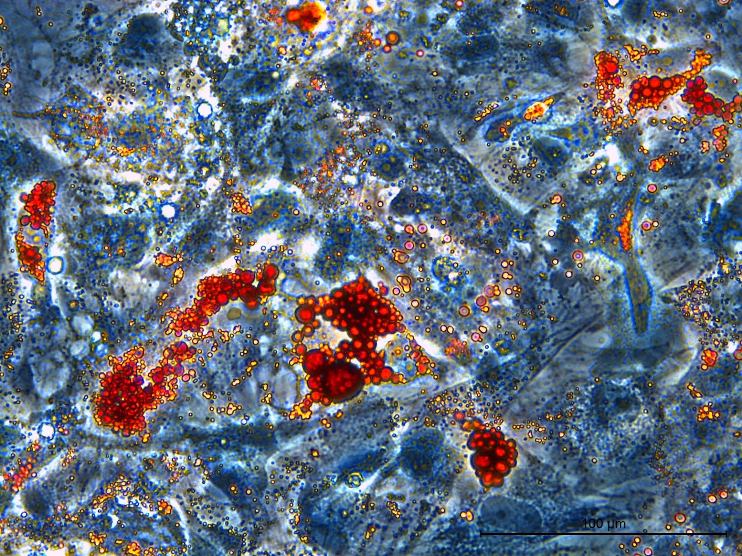
This picture shows cord tissue cells which have been turned into adipocytes (fat-forming cells).
To demonstrate that the differentiation has been successful, the cells are stained with a dye called oil red O.
The fat inside the cells appears as round droplets, which stain bright red.
To demonstrate that the cells can turn into chondrocytes (cartilage forming cells) we grow tiny (less than 1mm across) spherical pieces of what is essentially artificial cartilage.
To check that this has worked, we take slices of this tissue which are just 0.005mm thick and stain them with a variety of chemicals to show that chondrocyte formation has been successful.
The experiment illustrated below shows cord tissue cells which have been turned into chondrocytes.
The first two images are of the same sample taken at two different magnifications. This sample has been stained with two different dyes:
• Nuclear fast red. This shows up the cells, which appear as irregular red spots.
• Alcian blue. This stains a class of molecules called glycosaminoglycans which are an important component of cartilage – they contribute to its shock-absorbing properties.


The next two images are the of same sample taken at two different magnifications.
This sample has been stained with safranin-O, a dye which binds to a class of molecules called proteoglycans which are also a major component of cartilage tissue.

